Web cookies (also called HTTP cookies, browser cookies, or simply cookies) are small pieces of data that websites store on your device (computer, phone, etc.) through your web browser. They are used to remember information about you and your interactions with the site.
Purpose of Cookies:
Session Management:
Keeping you logged in
Remembering items in a shopping cart
Saving language or theme preferences
Personalization:
Tailoring content or ads based on your previous activity
Tracking & Analytics:
Monitoring browsing behavior for analytics or marketing purposes
Types of Cookies:
Session Cookies:
Temporary; deleted when you close your browser
Used for things like keeping you logged in during a single session
Persistent Cookies:
Stored on your device until they expire or are manually deleted
Used for remembering login credentials, settings, etc.
First-Party Cookies:
Set by the website you're visiting directly
Third-Party Cookies:
Set by other domains (usually advertisers) embedded in the website
Commonly used for tracking across multiple sites
Authentication cookies are a special type of web cookie used to identify and verify a user after they log in to a website or web application.
What They Do:
Once you log in to a site, the server creates an authentication cookie and sends it to your browser. This cookie:
Proves to the website that you're logged in
Prevents you from having to log in again on every page you visit
Can persist across sessions if you select "Remember me"
What's Inside an Authentication Cookie?
Typically, it contains:
A unique session ID (not your actual password)
Optional metadata (e.g., expiration time, security flags)
Analytics cookies are cookies used to collect data about how visitors interact with a website. Their primary purpose is to help website owners understand and improve user experience by analyzing things like:
How users navigate the site
Which pages are most/least visited
How long users stay on each page
What device, browser, or location the user is from
What They Track:
Some examples of data analytics cookies may collect:
Page views and time spent on pages
Click paths (how users move from page to page)
Bounce rate (users who leave without interacting)
User demographics (location, language, device)
Referring websites (how users arrived at the site)
Here’s how you can disable cookies in common browsers:
1. Google Chrome
Open Chrome and click the three vertical dots in the top-right corner.
Go to Settings > Privacy and security > Cookies and other site data.
Choose your preferred option:
Block all cookies (not recommended, can break most websites).
Block third-party cookies (can block ads and tracking cookies).
2. Mozilla Firefox
Open Firefox and click the three horizontal lines in the top-right corner.
Go to Settings > Privacy & Security.
Under the Enhanced Tracking Protection section, choose Strict to block most cookies or Custom to manually choose which cookies to block.
3. Safari
Open Safari and click Safari in the top-left corner of the screen.
Go to Preferences > Privacy.
Check Block all cookies to stop all cookies, or select options to block third-party cookies.
4. Microsoft Edge
Open Edge and click the three horizontal dots in the top-right corner.
Go to Settings > Privacy, search, and services > Cookies and site permissions.
Select your cookie settings from there, including blocking all cookies or blocking third-party cookies.
5. On Mobile (iOS/Android)
For Safari on iOS: Go to Settings > Safari > Privacy & Security > Block All Cookies.
For Chrome on Android: Open the app, tap the three dots, go to Settings > Privacy and security > Cookies.
Be Aware:
Disabling cookies can make your online experience more difficult. Some websites may not load properly, or you may be logged out frequently. Also, certain features may not work as expected.
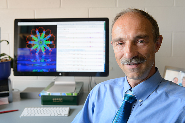
A Channel 8 science story about 3D printing called upon the expertise of UConn’s Dr. Anson Ma to explain some of the current and potential benefits of the technology.
Dr. Ma had challenged his students to create an artificial kidney using 3D technology and they produced it using 3D printing.
“Right now this is a prototype,” said Ma. “The longer term goal is can we incorporate cells, for example stem cells from the patients, then we can create fully compatible organs.”
The story highlights UConn’s commitment to new and emerging technologies as well as the ever-growing importance of materials science.
This self-assembling protein nanoparticle relies on rigid protein structures called ‘coiled coils’ (blue and green in the image) to create a stable framework upon which scientists can attach malaria parasite antigens. Early tests show that injecting the nanoparticles into the body as a vaccine initiates a strong immune system response that destroys a malarial parasite when it enters the body and before it has time to spread. (Image courtesy of Peter Burkhard)
A self-assembling nanoparticle designed by a UConn professor is the key component of a potent new malaria vaccine that is showing promise in early tests.
For years, scientists trying to develop a malaria vaccine have been stymied by the malaria parasite’s ability to transform itself and “hide” in the liver and red blood cells of an infected person to avoid detection by the immune system.
But a novel protein nanoparticle developed by Peter Burkhard, a professor in the Department of Molecular & Cell Biology, in collaboration with David Lanar, an infectious disease specialist with the Walter Reed Army Institute of Research, has shown to be effective at getting the immune system to attack the most lethal species of malaria parasite, Plasmodium falciparum, after it enters the body and before it has a chance to hide and aggressively spread.
The key to the vaccine’s success lies in the nanoparticle’s perfect icosahedral symmetry (think of the pattern on a soccer ball) and ability to carry on its surface up to 60 copies of the parasite’s protein. The proteins are arranged in a dense, carefully constructed cluster that the immune system perceives as a threat, prompting it to release large amounts of antibodies that can attack and kill the parasite.
In tests with mice, the vaccine was 90-100 percent effective in eradicating the Plasmodium falciparum parasite and maintaining long-term immunity over 15 months. That success rate is considerably higher than the reported success rate for RTS,S, the world’s most advanced malaria vaccine candidate currently undergoing phase 3 clinical trials, which is the last stage of testing before licensing.
Peter Burkhard, professor of molecular and cell biology, with an image of the protein nanoparticle he designed. (Peter Morenus/UConn Photo)
“Both vaccines are similar, it’s just that the density of the RTS,S protein displays is much lower than ours,” says Burkhard. “The homogeneity of our vaccine is much higher, which produces a stronger immune system response. That is why we are confident that ours will be an improvement.
“Every single protein chain that forms our particle displays one of the pathogen’s protein molecules that are recognized by the immune system,” adds Burkhard, an expert in structural biology affiliated with UConn’s Institute of Materials Science. “With RTS,S, only about 14 percent of the vaccine’s protein is from the malaria parasite. We are able to achieve our high density because of the design of the nanoparticle, which we control.”
The search for a malaria vaccine is one of the most important research projects in global public health. The disease is commonly transported through the bites of nighttime mosquitoes. Those infected suffer from severe fevers, chills, and a flu-like illness. In severe cases, malaria causes seizures, severe anemia, respiratory distress, and kidney failure. Each year, more than 200 million cases of malaria are reported worldwide. The World Health Organization estimated that 627,000 people died from malaria in 2012, many of them children living in sub-Saharan Africa.
It took the researchers more than 10 years to finalize the precise assembly of the nanoparticle as the critical carrier of the vaccine and find the right parts of the malaria protein to trigger an effective immune response. The research was further complicated by the fact that the malaria parasite that impacts mice used in lab tests is structurally different from the one infecting humans.
The scientists used a creative approach to get around the problem.
Infectious disease specialist David Lanar of the Walter Reed Army Institute of Research, who is collaborating with UConn professor Peter Burkhard in pursuing a new malaria vaccine. (Photo courtesy of David Lanar)
“Testing the vaccine’s efficacy was difficult because the parasite that causes malaria in humans only grows in humans,” Lanar says. “But we developed a little trick. We took a mouse malaria parasite and put in its DNA a piece of DNA from the human malaria parasite that we wanted our vaccine to attack. That allowed us to conduct inexpensive mouse studies to test the vaccine before going to expensive human trials.”
The pair’s research has been supported by a $2 million grant from the National Institutes of Health and $2 million from the U.S. Military Infectious Disease Research Program. A request for an additional $7 million in funding from the U.S. Army to conduct the next phase of vaccine development, including manufacturing and human trials, is pending.
“We are on schedule to manufacture the vaccine for human use early next year,” says Lanar. “It will take about six months to finish quality control and toxicology studies on the final product and get permission from the FDA to do human trials.”
Lanar says the team hopes to begin early testing in humans in 2016 and, if the results are promising, field trials in malaria endemic areas will follow in 2017. The required field trial testing could last five years or more before the vaccine is available for licensure and public use, Lanar says.
Martin Edlund, CEO of Malaria No More, a New York-based nonprofit focused on fighting deaths from malaria, says, “This research presents a promising new approach to developing a malaria vaccine. Innovative work such as what’s being done at the University of Connecticut puts us closer than we’ve ever been to ending one of the world’s oldest, costliest, and deadliest diseases.”
A Switzerland-based company, Alpha-O-Peptides, founded by Burkhard, holds the patent on the self-assembling nanoparticle used in the malaria vaccine. Burkhard is also exploring other potential uses for the nanoparticle, including a vaccine that will fight animal flu and one that will help people with nicotine addiction. Professor Mazhar Khan from UConn’s Department of Pathobiology is collaborating with Burkhard on the animal flu vaccine.
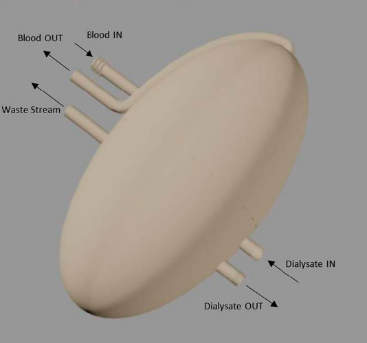
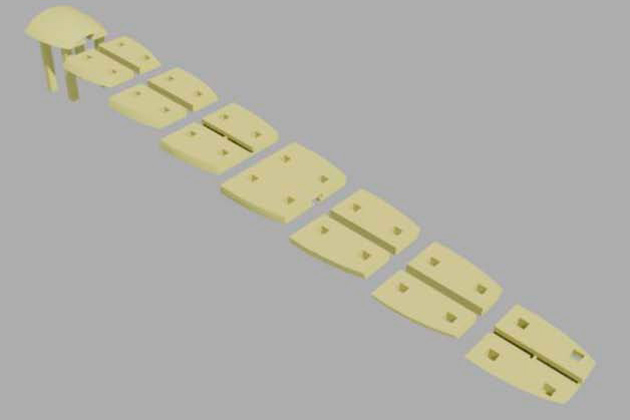
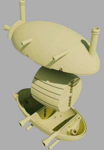
Senior chemical engineering student Derek Chhiv, right, discusses with Professor Anson Ma his group’s prototype for an artificial kidney. The prototype was generated through 3-D printing. (Al Ferreira for UConn)Three-dimensional printing has garnered coverage in the popular press for its application in the custom manufacturing of tools and mechanical parts. But six School of Engineering seniors have recently taken the application of the technology into the medical field, using 3-D printing to create body parts.Under the direction of Anson Ma, assistant professor in the Department of Chemical and Biomolecular Engineering and the Institute of Materials Science, two three-person teams of chemical engineering students were tasked with creating an artificial kidney for their Senior Design Project using 3-D printing technology. 3-D printing is an additive manufacturing method capable of creating complex parts that are otherwise impossible or extremely difficult to produce.The students participating were: Derek Chhiv, Meaghan Sullivan, Danny Ung, Benjamin Coscia, Guleid Awale, and Ali Rogers. They are one of the first classes of students to partner with a commercial 3-D printing company, ACT Group, to create a prototype.A drawing of the shell of an artificial kidney rendered using AutoCAD software. It is 12 cm long and 6 cm in diameter, an average size for an adult human biological kidney. (Image courtesy of Benjamin Coscia ’14 (ENG))The challenge the teams set out to tackle is rooted in a very real problem.The United States Renal Data System reports that, as recently as 2009, End-Stage Renal Disease (ESRD) resulted in over 90,000 deaths. Options for treatment of renal disease are essentially limited to either an organ transplant or dialysis. However, there is a limited supply of transplantable kidneys, with demand far outstripping the supply; and dialysis is expensive and is only a temporary solution.According to data from the National Kidney Foundation, there are currently nearly 100,000 people awaiting kidney transplants in the United States, yet only 14,000 kidney transplants took place in the country this year. An additional 2,500 new patients are added to the kidney waiting list each month.Faced with these challenges, the two UConn teams set out on a year-long effort to design and develop a prototype of a cost-effective, functional artificial kidney using chemical engineering principles and 3-D printing technology.“The objective of the design project is to get these students to combine the latest technology and their chemical engineering knowledge, learned over their four years at UConn, to solve a technical problem where we can make a difference,” notes Ma. “Can they push the technology further?”Guleid Awale, one of the seniors, said the two design teams each took a slightly different approach to the problem. “While the other team utilized techniques such as electrodialysis and forward osmosis in their prototype, our group opted for mainly hollow fiber membrane technology commonly found in traditional hemodialysis treatments.”Benjamin Coscia ’14 (ENG) explains the hollow fiber membrane technology: “Because 3D printing resolutions are not currently low enough to print a structure which will actually filter blood, the file is of only the shell of the kidney. Hollow fiber membranes will be installed on the inside to do the filtration function. The kidney will then be sealed together using the threads and sealing o-rings. A fluid called dialysate will be circulated on the outside of the membranes, inside of the shell, which will cause flux of components from the blood. A waste stream maintains the person’s ability to urinate. The outside of the shell can be used as a substrate for growth of biological material for ease of integration into the body.”The outer shell of the artificial kidney prototype using electrodialysis and forward osmosis.Assembling the components of the 3-D kidney. (Image courtesy of Derek Chhiv ’14 (ENG)) Interior components of the 3-D artificial kidney, rendered using AutoCAD software.Assembling the kidney components. (Images courtesy of Derek Chhiv ’14 (ENG)) After undertaking the research and development of the design, the teams designed the prototype using AutoCAD software. Then each team collaborated with UConn technology partner ACT Group of Cromwell, Conn. to select the appropriate polymers, as well as the right printer to use in printing the particular prototype design.The two teams presented their projects on May 2 at the School of Engineering Senior Design Demonstration Day.“The biggest challenge in approaching the project was applying the engineering knowledge we’ve gained during our undergraduate years to a more complex biological application,” Awale notes. “This forced us to come out of our comfort zone and rely on our problem-solving skills in order to come up with viable solutions.”
Professor Sanjeeva Murthy from Rutgers University, the Polymer Program Seminar speaker on March 28th, is an alum of the program and IMS. He earned his Ph.D. in Materials Science in 1976 under Jim Knox (MCB), one of the founding members of the Polymer Program, who retired in 2002. Sanjeeva Murthy was one of the first graduates of IMS. Jim Knox was able to show off the champagne bottle from Sanjeeva’s Ph.D. defense celebration, although he later admitted that Sanjeeva had probably not taken a single sip from it!
During his lecture, Sanjeeva mentioned that, when he started his PhD studies here over 40 years ago in 1972, the IMS building was new, there was no Storrs Downtown or even the NCAA basketball bracket!
After his seminar Sanjeeva said, “‘I had a great time today. It brought back all memories of years ago. I also enjoyed my discussions with the new faculty. I hope to be in touch with some of them with whom I have common interest. I thank you all for inviting me today. Look forward to seeing you again.”
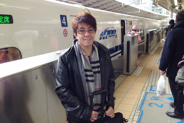
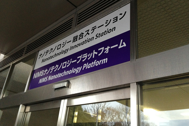
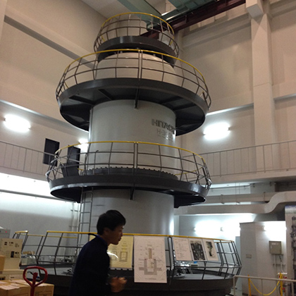
Professor Anson Ma takes the Shinkansen (high-speed train) from Tokyo to Kyoto, during a five-day trip to Japan with the Young Scientist Exchange. (Photos courtesy of Anson Ma)UConn researcher Anson Ma recently participated in a prestigious U.S.-Japan Young Scientist Exchange Program that enabled him to spend five days visiting top Japanese universities and research centers, where he presented his research on rheology and processing of nanofluids and met with fellow young researchers.The National Science Foundation (NSF) and the Ministry of Education, Culture, Sports, Science, & Technology in Japan (MEXT) initiated the science diplomacy-style exchange program in 2003 to foster collaborations among U.S. and Japanese researchers in strategic areas. Leading young Japanese academics visit U.S. universities and researchers, and U.S. academics reciprocate.The trip included a visit to the National Institute for Materials Science, including laboratories associated with the institute’s nanotechnology platform.Ma, an assistant professor of chemical and biomolecular engineering, was nominated by a senior researcher for his contributions in understanding the flow behavior and processing of complex fluids for biomedical and energy applications. During day-long workshops from Dec. 9 to 13, Ma and his fellow U.S. and Japanese scientists delivered and attended presentations, toured laboratories, and discussed avenues for collaboration.The group visited the National Institute for Materials Science, including laboratories associated with the institute’s nanotechnology platform; the University of Tokyo; Osaka University; and Kyoto University. Among the technology highlights that particularly impressed Ma was the remarkable ultra-high voltage Hitachi electron microscope housed at Osaka University, which is more than 13 meters high.The ultra-high voltage electron microscope at Osaka University.The delegates also enjoyed one day of sightseeing, when they took the high-speed Shinkansen train (also known as the ‘bullet train’) from Tokyo to Kyoto for a tour of the 17th-century Kodaiji Temple.The research trip was organized and led by Alexander Revzin, currently a program director in the Biosensing Division at the National Science Foundation and a University of California-Davis professor, and Dino Di Carlo, associate professor of bioengineering at UCLA.“The goal is to unveil areas of mutual interest and to build collaborative research bridges in transformative research arenas,” says Di Carlo.
Professor Hidetoshi Kotera, executive vice-president of Kyoto University for external strategy, knowledge, and technology transfer and innovation, speaks about current research activities and future plans for the university.The exchange program focuses on bio-nano-micro technologies, and while the themes have remained constant since 2003, the application areas – for example, manufacturing, sensing, and energy – of the visits vary from year to year. When Japanese delegates come to the U.S., they visit various different U.S. universities during their exchange tours; in recent years, these have included UCLA, Caltech, MIT, Harvard, Northwestern, and the University of North Carolina.Ma says the experience was extremely worthwhile, noting that he met potential collaborators among the U.S. delegates as well as among the Japanese faculty. He found the work of three Japanese researchers particularly compelling. One is involved in biomechanics research focusing on the motion of cells, and another is developing a bioadhesive for creating 3-D tissue using cells as building blocks – “just like playing with Lego blocks,” says Ma. A third is developing advanced biomimetic materials.
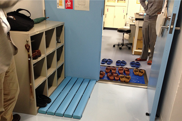
A visit to Tokyo University, where shoes are not allowed in many labs; visitors must exchange their shoes for slippers.Ma was also impressed with the laboratories and cleanroom facilities, which he says were organized and efficient. However, he was surprised to find that in Japanese laboratories, as in living spaces, scientists must don slippers before entering research spaces – a custom that is forbidden in U.S. labs.Learn more about Ma’s research program here.
Dr. Douglas Adamson and Dr. Thomas Seery were awarded a NSF Grant of $200,000 for the project, “Unimolecular Micelles: Design, Synthesis, and Properties.” The grant was funded by the Macromolecular, Supramolecular and Nanochemistry Program of the Chemistry Division.
The project aims to synthesize and observe polymers that can create stable, single chain globules in solutions. Dr. Adamson says that “protein folds in such a way as to hide most of those insoluble amino acids while the leaving the water soluble ones near the surface.” The objective is to understand how artificial polymers can imitate the nanostructure forming abilities of proteins at a very fundamental level.
The formations of these accurately discrete structures are a continuous challenge for chemists. Adamson and Seery believe that the results of the project “will lead to applications such as robust artificial enzymes” and “plastic antibodies that function much like natural antibodies but avoid the need for biological source.” The morphology within these nanostructures can impact vast areas of technology such as medicines, electronics and biotechnology.
Now with the funding of NSF, Dr. Adamson and Dr. Seery are able to proceed in the process of exploring synthetic materials that may perform some of the functions of proteins. The project will also involve visits to local schools and will contribute to the training of undergraduate and graduate students.
Dr. Douglas H. Adamson received his B.S. degree at the University of Evansville, Indiana and his Ph.D. degree at University of Southern California. He joined the University of Connecticut in August 2008, becoming an Associate Professor in the Polymer Program at IMS with Chemistry as his home department. Dr. Adamson was appointed Director of the Polymer Program in July 2011.
Dr. Thomas Seery, Associate Professor of Chemistry, received his B.A. degree at Harvard University and his Ph. D. degree at University of Southern California. He joined the University of Connecticut in 1994. Dr. Seery’s research interests include studying polymer synthesis at surfaces and physical chemistry of polymers in solution.
Dr. Steven L. Suib, Board of Trustees Distinguished Professor of Chemistry, and recently appointed Director of IMS, has formed a revised Internal Advisory Board.
Since the conception of IMS, the assignment of the Internal Advisory Board has been to provide suggestions and solutions for problems of broad interest within IMS. The Internal Advisory Board consists of ten faculty members from five different departments. “We collaborate as a unit and lay out our vision for the general operation of IMS,” Dr. Ramamurthy Ramprasad, MSE Professor says. The feedback and ideas are forwarded to Director of IMS, Dr. Suib.
Dr. Suib was appointed the new Director of IMS on July 1st, 2013. The former Director of IMS, Dr. Harris Marcus, stepped down after 18 years of service. He will remain on the faculty in the Materials Science & Engineering Department.
The board remains the same throughout the academic year unless specific developments necessitate a change. “The board doesn’t change unless a member from the board resigns and is replaced, or if a member is removed,” says Deborah Perko, Executive Assistant of Infrastructure.
The current board members are:
Dr. Douglas H. Adamson: Director of the Polymer Program and Associate Professor of Chemistry
Dr. Mark Aindow: Associate Director of the Institute of Materials Science and Professor of Materials Science and Engineering
Dr. S. Pamir Alpay: Department Head and Professor of Materials Science and Engineering
Dr. A. Jon Goldberg: Professor of Oral Rehabilitation, Biomaterials and Skeletal Development at University of Connecticut Health Center
Dr. Faquir Jain: Professor of Electrical & Computer Engineering
Dr. Ramamurthy Ramprasad: Professor of Materials Science and Engineering
Dr. Thomas Seery: Associate Professor of Chemistry
Dr. Steven L. Suib: Director of IMS and Board of Trustees Distinguished Professor
Dr. Carolyn M. Teschke: Professor of Molecular & Cell Biology